The specimen height and eucentric height
What defines the position of specimen? Well, pretty
obviously, the specimen. But if we think about it, there's
nothing to stop us running the microscope with the specimen
moved up or down relative to the objective and condenser
lenses.
Suppose we moved the specimen up, as in the next Figure:
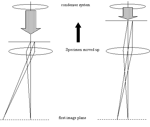
We can still focus on the specimen and make the microscope
work by slightly de-exciting (defocusing) the objective lens
and, if necessary, slightly increasing the excitation of the
condenser lens. In the picture we haven't worried deeply
about the exact arrangement of the illuminating coming out
of the condenser system because its not important in the
present discussion. All that's important is that we can
simply de-excite the objective and still form an image on
the first image plane, which then gets imaged onto the
phosphor screen. We have drawn four rays in both diagrams,
a pair from a point in the specimen on the optic axis, and a
pair from another point which off-axis. Note that the
magnification has changed slightly.
On most microscopes, we can also change the physical height
of the specimen. The height adjustment of the specimen is
sometimes called the 'eucentric height' adjustment or 'the z-
shift' or 'z-adjustment'. 'Eucentric' is a complicated word
for an easy idea described shortly. Z is 'z' because when
we move the specimen laterally we call it x-y shift, and so
'z' is as in 'coordinates x,y and z', meaning that z is a
vertical movement.
Ask the demonstrator: How do I mechanically adjust the eucentric
height or, in other words, the specimen z shift?
You will be shown a mechanical adjustment on the specimen
holder, although many modern machines have an electrical servo
drive for this adjustment.
Experiment: In image mode, form an in-focus image of the
specimen. Make sure the step size of the objective lens is
at a high setting. Adjust the z-shift. See what happens to
the image. Refocus the image using the objective lens (ie
the focus knob). By noting which way you have to turn the
focus knob, work out whether the specimen has moved up or
down. Repeat the experiment until you
have a feeling for how a movement in z corresponds to a
change in focus of the objective lens. Can you notice any
change in magnification as you change the objective focus?
Ask the demonstrator: How do I tilt the specimen?
You may be shown a mechanical knob on the specimen holder,
an electrical device, or possibly one or two foot pedals.
These all have the effect, either directly, or via a motor,
of rotating the specimen holder so that the specimen tilts
over, away from the horizontal.
Experiment: Try tilting the specimen back forth. Watch the
image to see if it moves. Change the z shift as before and
repeat the experiment. Can you work out what's happening?
You should be able to find an adjustment of the z-shift
where tilting the specimen leads to a minimal movement of
the image. This is called the eucentric height, which means
'the height of the specimen at which its image does not
moved laterally as a function of specimen tilt'.
What happens is this. Unless you are using an unusual type
of microscope which has a 'top entry stage', then the
specimen is supported on a thin rod which comes in
horizontally from the outside of the objective lens (a 'side
entry stage'). The rod can rotate around a fixed axis,
tilting the specimen. The z-shift adjustment effectively
lifts or lowers the specimen without affecting the rotation
axis of the rod. (Exactly how it does this varies according
to the design of the specimen holder).
It is common practice to adjust the z-shift so that the
middle of the specimen lies on the rotation axis of the
specimen loading arm. This has the convenience of meaning
that when the specimen is tilted, the point you are
observing remains stationary: i.e. we adjust the specimen to
the eucentric height.
All sorts of aspects of the performance of the microscope
depend upon the exact height of the specimen. The height of
the specimen defines the excitation of the objective lens,
which affects the magnification of the whole
machine. There is a further complication that we will learn
about in more detail later: the magnetic field of the
objective lens actually spills over the top of the specimen.
This means the objective setting also affects the
illumination beam, in a way which is determined by the
specimen height, and which is very important when we come on
to STEM imaging. The resolution of the microscope is wholly
determined by the performance of the objective, and hence
also by the specimen height.
For this reason, the
manufacturer will often recommend that the microscope should
always be run with the specimen at the eucentric height. In
fact, you may find you can improve the performance of the
microscope for certain applications by operating at a
different specimen heights. Many microscopists
determine the very best setting of the objective lens for a
particular application (say, high-resolution imaging) and
then adjust the specimen height to get the microscope in
focus, even if this means the specimen is non-eucentric.


Copyright J M Rodenburg
|