The twin objective lens
We now cover a couple of subjects in more detail, starting with
the heart of the electron microscope, the objective lens.
Everything we learnt in the earlier sections about the objective
lens is only half the story. As a model in your mind for
how to line it up, it is an accurate and
useful picture. However, the whole truth is somewhat more
complicated.
A modern electron microscope has many different and
elaborate 'modes', including STEM mode, which we will cover later. In order to accommodate all these
different operations, lens design has become a subtle art.
We hinted earlier that some of the objective lens 'leaks
over' the specimen, and so changes the focus of the beam
before it hits the sample. In fact, this is a slight
understatement. In a TEM/STEM machine, the objective lens
pre-field can be as strong as the lower half of the
objective lens. It depends on what type of microscope you
are using. Look at the schematic diagram again. Is half
the objective lens above the specimen? Ask the demonstrator if
in doubt. A symmetric objective lens, which is now very
common, has as much lens above as below the specimen.
A symmetric objective has an important consequence: the
first (upper) half of the objective lens acts like a third
condenser lens, and so in truth there are two beam cross-
overs between C2 and the specimen, as shown in the next
diagram. (Note that the exact details of the objective pre-field can
be affected a mini-lens mounted above the specimen: this varies
between makes of microscope, but the general principles are the same.)
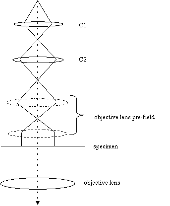
Notice that the objective lens pre-field actually does two
things: it forms the second cross-over below C2 and then
can be used to make the beam parallel before it hits the
specimen.
All this may seem to make things appear much more
complicated. However, if you think about it, the behaviour
of the cross-over at the specimen (i.e. the first experiment
we ever did changing C2), goes ahead as before, except
there is now one other cross-over. As C2 is lowered in
strength, both cross-overs descend in the column, until the
second cross-over is a sharp point on the specimen plane,
focussed through the lower part of the objective pre-field,
and therefore forming an image of the filament. Ray
diagrams for increasing values of C2 are shown schematically
below:

Assume that the horizontal dotted line is at the focal
length of the lower part of the objective lens pre-field: we
can tell this from the right-most diagram, where the
illumination in the specimen place is parallel and hence, by
definition, the previous cross-over is at the focal length .
In the left-most diagram, C2 is low, and the beams are
entering the top of the objective parallel. The beam cross-
over between the two parts of the pre-field lens is slightly
higher than in the normal setting (right-most diagram), and
so the beam on the specimen is slightly convergent. As C2
becomes more excited, the first cross-over passes through
the top part of the pre-field: remember that rays through
the centre of a lens pass through it in a straight line. At
some point, a second cross-over is formed at the specimen
plane (which is when we see the filament in focus), which in
turn passes up through lower part of the pre-field.
Of course in reality, the objective pre-field is one
continuous lens, and so it is quite wrong to think of
straight rays passing through two discrete lens, but the net
effect is the same. To all intents and purposes, thinking of a single objective
mounted below the specimen is conceptually equivalent to the to what
happens with a twin objective, except that there is an extra
cross-over, and at the final setting the illuminating beam
is parallel, not divergent.
As far as alignment of the condenser system and objective is
concerned, everything we said in the previous chapters still
applies.
Remember: the system is usually designed in
such a way that the illumination is parallel at the specimen
when the intensity (C2) knob is high.


Copyright J M Rodenburg
|